Well, I'm not sure how the formatting is going to turn out on this article, I hope it is readable! It sure is interesting.
Dr. Helise Coopersmith, North Shore-LIJ Health System
Date: 02 July 2013 Time: 06:58 PM ET
Dr. Helise Coopersmith is a musculoskeletal and body imaging radiologist for the North Shore-LIJ Health System, assistant professor of radiology at the Hofstra North Shore-LIJ School of Medicine and a member of the Hofstra medical school's admissions committee. She contributed this article to LiveScience's Expert Voices: Op-Ed & Insights.
I have worked as a musculoskeletal radiologist for many years and have seen a wide range of bone injuries. But recently, I found myself for the first time using my X-ray table to look at a 2,500-year-old bone and a piece of an ancient arrow.
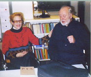
The bone, discovered in Northern Greece, was brought to me by Anagnostis Agelarakis, a professor and chair of anthropology at Adelphi University. It was a section of the ulna bone, which is the inner of two forearm bones.
My initial impression was surprise. Although the outer region of bone, the cortex, was thinned by time, and the inner region, the medullary cavity, had long since disintegrated, the girth and contours of the bone were quite similar to a human bone one would see today.
Beside my X-ray table, I had a photograph of the re-assembled skull that was found with the ulna bone and a sketch by scientific illustrator Argie Agelarakis (Anagnostis's wife) of what the soldier's face may have looked like around the time of his eventual death, presumably at about 58 to 62 years of age.
My team and I took three X-rays of the ulna bone, and we found that indeed the films confirmed what Anagnostis Agelarakis had suspected.
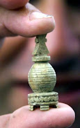
There was a barbed component to the arrowhead that could not be seen with the naked eye. The full extent of the remaining arrowhead could now be seen and was seated superficially within the bone, located only within the cortex, or outer shell. This supported Agelarakis's notion that the arrowhead could have been removed if not for its barbed component.
There was a large bony (osseous) spur adjacent to the arrowhead, which make sense as the human body can form extra bone material in response to trauma. Such spurs take many months to fully mature, which implies that the soldier lived for a long time after the injury. Also, there was no bony erosion adjacent to the arrowhead, confirming that the arrowhead did not cause life-threatening infection.
We also noted that the arrowhead and the osseous spur were in the region of the flexor digitorum profundus muscle, which means the injury would have made it difficult for the soldier to flex his fingers and grasp objects.
There was a story behind the objects we were seeing, the story of an ancient Greek warrior who was an injured veteran, like many who are celebrated today and served by the hospital I work in.
It is amazing to think that the same X-ray technology that we use to diagnose conditions for our patients can answer age-old questions and help solve historical mysteries.


No comments:
Post a Comment